Explore
 Validate
 Learn
Learn ELISA
ELISA Immunocytochemistry
Antibody data
- Antibody Data
- Antigen structure
- References [2]
- Comments [0]
- Validations
- Immunocytochemistry [4]
- Immunohistochemistry [3]
- Other assay [3]
Submit
Validation data
Reference
Comment
Report error
- Product number
- ABS 028-08-02 - Provider product page

- Provider
- Invitrogen Antibodies
- Product name
- NPY Monoclonal Antibody (8)
- Antibody type
- Monoclonal
- Antigen
- Synthetic peptide
- Description
- ABS 028-08-02 detects Neuropeptide Y in human and rat samples. ABS 028-08-02 has been successfully used in ELISA and immunohistochemistry (paraffin) procedures. NOTE: Concentration is lot-dependent and can vary from 0.85-1.15 mg/mL
- Reactivity
- Human, Mouse, Rat
- Host
- Mouse
- Isotype
- IgG
- Antibody clone number
- 8
- Vial size
- 400 μL
- Concentration
- 1.0 mg/mL
- Storage
- 4°C, store in dark
Submitted references Endometritis Changes the Neurochemical Characteristics of the Caudal Mesenteric Ganglion Neurons Supplying the Gilt Uterus.
Ghrelin Regulates Glucose and Glutamate Transporters in Hypothalamic Astrocytes.
Jana B, Całka J
Animals : an open access journal from MDPI 2020 May 20;10(5)
Animals : an open access journal from MDPI 2020 May 20;10(5)
Ghrelin Regulates Glucose and Glutamate Transporters in Hypothalamic Astrocytes.
Fuente-Martín E, García-Cáceres C, Argente-Arizón P, Díaz F, Granado M, Freire-Regatillo A, Castro-González D, Ceballos ML, Frago LM, Dickson SL, Argente J, Chowen JA
Scientific reports 2016 Mar 30;6:23673
Scientific reports 2016 Mar 30;6:23673
No comments: Submit comment
Supportive validation
- Submitted by
- Invitrogen Antibodies (provider)
- Main image
- Experimental details
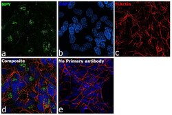
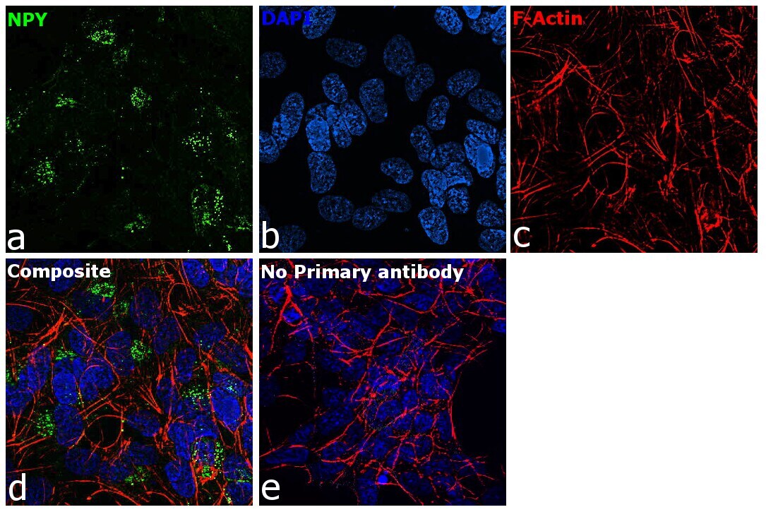
- Immunofluorescence analysis of NPY was performed using 70% confluent log phase SH-SY5Y cells. The cells were fixed with 4% paraformaldehyde for 10 minutes, permeabilized with 0.1% Triton™ X-100 for 15 minutes, and blocked with 2% BSA for 1 hour at room temperature. The cells were labeled with NPY Monoclonal Antibody (8) (Product # ABS 028-08-02) at 5 µg/mL in 0.1% BSA, incubated at 4 degree celsius overnight and then labeled with Donkey anti-Mouse IgG (H+L) Highly Cross-Adsorbed Secondary Antibody, Alexa Fluor Plus 488 (Product # A32766), (1:2000), for 45 minutes at room temperature (Panel a: Green). Nuclei (Panel b:Blue) were stained with ProLong™ Diamond Antifade Mountant with DAPI (Product # P36962). F-actin (Panel c: Red) was stained with Rhodamine Phalloidin (Product # R415, 1:300). Panel d represents the merged image showing cytoplasmic(Golgi complex like pattern) localization. Panel e represents control cells with no primary antibody to assess background. The images were captured at 60X magnification.

- Submitted by
- Invitrogen Antibodies (provider)
- Main image

- Experimental details
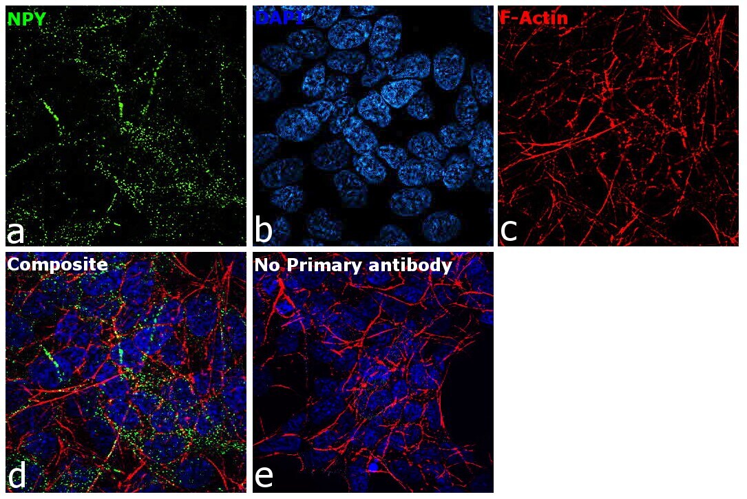
- Immunofluorescence analysis of NPY was performed using 70% confluent log phase SH-SY5Y cells. The cells were fixed with 4% paraformaldehyde for 10 minutes, permeabilized with 0.1% Triton™ X-100 for 15 minutes, and blocked with 2% BSA for 1 hour at room temperature. The cells were labeled with NPY Monoclonal Antibody (8) (Product # ABS 028-08-02) at 5 µg/mL in 0.1% BSA, incubated at 4 degree celsius overnight and then labeled with Donkey anti-Mouse IgG (H+L) Highly Cross-Adsorbed Secondary Antibody, Alexa Fluor Plus 488 (Product # A32766), (1:2,000), for 45 minutes at room temperature (Panel a: Green). Nuclei (Panel b:Blue) were stained with ProLong™ Diamond Antifade Mountant with DAPI (Product # P36962). F-actin (Panel c: Red) was stained with Rhodamine Phalloidin (Product # R415, 1:300). Panel d represents the merged image showing cytoplasmic (vesicular like pattern) localization. Panel e represents control cells with no primary antibody to assess background. The images were captured at 60X magnification.
- Submitted by
- Invitrogen Antibodies (provider)
- Main image
- Experimental details
- Immunofluorescence analysis of NPY was performed using 70% confluent log phase SH-SY5Y cells. The cells were fixed with 4% paraformaldehyde for 10 minutes, permeabilized with 0.1% Triton™ X-100 for 15 minutes, and blocked with 2% BSA for 1 hour at room temperature. The cells were labeled with NPY Monoclonal Antibody (8) (Product # ABS 028-08-02) at 5 µg/mL in 0.1% BSA, incubated at 4 degree celsius overnight and then labeled with Donkey anti-Mouse IgG (H+L) Highly Cross-Adsorbed Secondary Antibody, Alexa Fluor Plus 488 (Product # A32766), (1:2,000), for 45 minutes at room temperature (Panel a: Green). Nuclei (Panel b:Blue) were stained with ProLong™ Diamond Antifade Mountant with DAPI (Product # P36962). F-actin (Panel c: Red) was stained with Rhodamine Phalloidin (Product # R415, 1:300). Panel d represents the merged image showing cytoplasmic (vesicular like pattern) localization. Panel e represents control cells with no primary antibody to assess background. The images were captured at 60X magnification.

- Submitted by
- Invitrogen Antibodies (provider)
- Main image

- Experimental details
- Immunofluorescence analysis of NPY was performed using 70% confluent log phase SH-SY5Y cells. The cells were fixed with 4% paraformaldehyde for 10 minutes, permeabilized with 0.1% Triton™ X-100 for 15 minutes, and blocked with 2% BSA for 1 hour at room temperature. The cells were labeled with NPY Monoclonal Antibody (8) (Product # ABS 028-08-02) at 5 µg/mL in 0.1% BSA, incubated at 4 degree celsius overnight and then labeled with Donkey anti-Mouse IgG (H+L) Highly Cross-Adsorbed Secondary Antibody, Alexa Fluor Plus 488 (Product # A32766), (1:2000), for 45 minutes at room temperature (Panel a: Green). Nuclei (Panel b:Blue) were stained with ProLong™ Diamond Antifade Mountant with DAPI (Product # P36962). F-actin (Panel c: Red) was stained with Rhodamine Phalloidin (Product # R415, 1:300). Panel d represents the merged image showing cytoplasmic(Golgi complex like pattern) localization. Panel e represents control cells with no primary antibody to assess background. The images were captured at 60X magnification.
Supportive validation
- Submitted by
- Invitrogen Antibodies (provider)
- Main image
- Experimental details
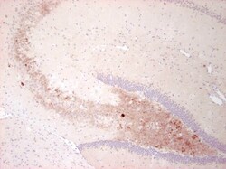
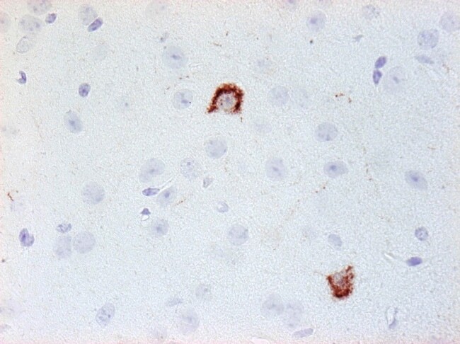
- Immunohistochemical analysis of Neuropeptide Y in paraffin-embedded mouse brain tissue (fibers in the hilus of the hippocampus) from a mouse suffering an epiletic attack using a Neuropeptide Y monoclonal antibody (Product # ABS 028-08-02) at a dilution of 1:1000.
- Submitted by
- Invitrogen Antibodies (provider)
- Main image

- Experimental details
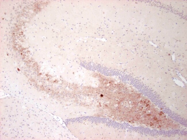
- Immunohistochemical analysis of Neuropeptide Y in enlarged section of a paraffin-embedded mouse brain tissue (fibers in the hilus of the hippocampus) from a mouse suffering an epileptic attack using a Neuropeptide Y monoclonal antibody (Product # ABS 028-08-02) at a dilution of 1:1000.
- Submitted by
- Invitrogen Antibodies (provider)
- Main image

- Experimental details
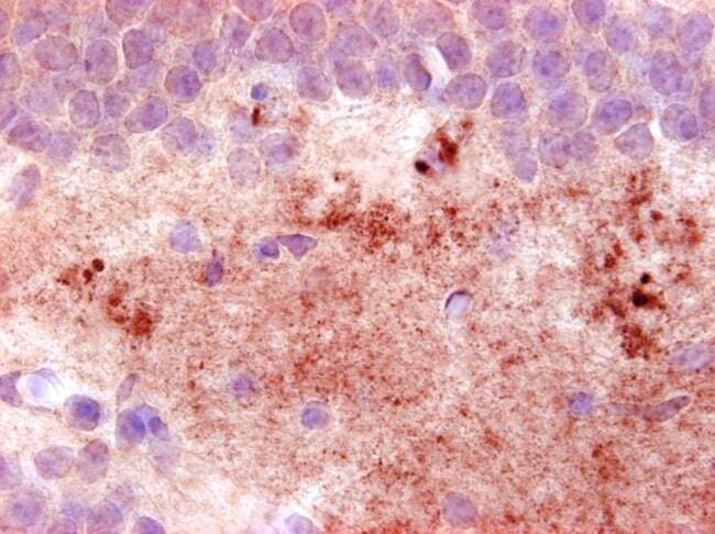
- Immunohistochemical analysis of Neuropeptide Y in paraffin-embedded mouse brain tissue (interneurons in the cortex) using a Neuropeptide Y monoclonal antibody (Product # ABS 028-08-02) at a dilution of 1:1000.
Supportive validation
- Submitted by
- Invitrogen Antibodies (provider)
- Main image
- Experimental details
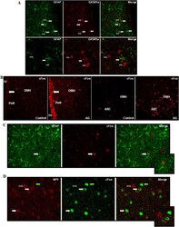
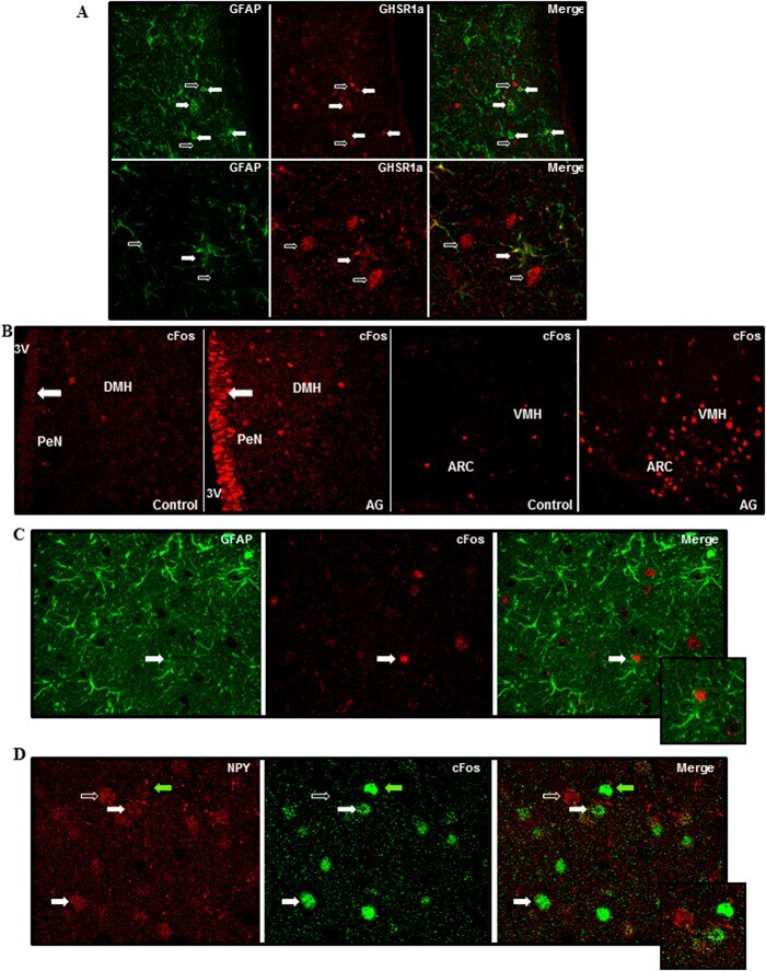
- Figure 1 Analysis of hypothalamic glial response to intracerebroventricular acyl-ghrelin treatment. ( A ) Double immunohistochemistry for glial fibrillary acidic protein (GFAP; green) and ghrelin receptor (GHSR1a; red) in the hypothalamus. Solid arrows indicate cells positive for both GFAP and GHSR1a. Open arrows indicate cells that are immunopositive only for GHSR1a. ( B ) Immunohistochemistry for cFos in control rats and rats that were treated icv with acyl-ghrelin (AG) for 1 hour. There was a clear increase in cFos positive cells (red) in the hypothalamus and in cells lining the 3 rd ventricle (3 V) after AG treatment (solid arrow). ( C ) Double immunohistochemistry for GFAP and cFos. Solid arrow indicates a cell that is immunopositive for both GFAP and cFos. ( D ) Double immunohistochemistry for neuropeptide Y (NPY) and cFos in the arcuate nucleus. Solid errors indicate double labeled cells and the open arrow indicates a NPY neuron that is not positive for cFos. The green arrow indicates a cell only positive for cFOS. ARC = arcuate nucleus, DMH = dorsomedial hypothalamus, PeN = periventricular nucleus, VMH = Ventromedial nucleus.
- Submitted by
- Invitrogen Antibodies (provider)
- Main image
- Experimental details
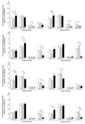
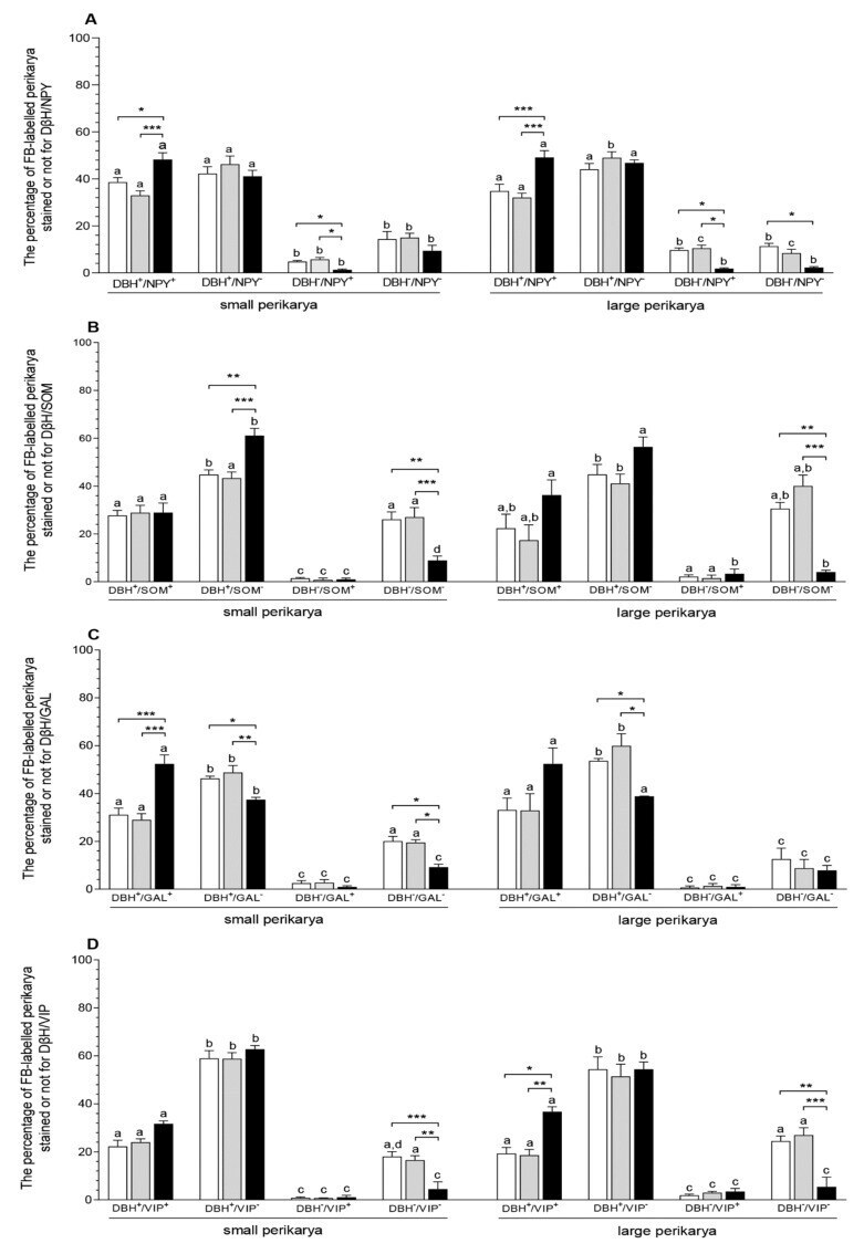
- Figure 2 The populations (expressed as percentages, mean +- SEM) of small and large uterine perikarya expressing dopamine-beta-hydroxylase (DbetaH) and/or neuropeptide Y (NPY) ( A ), DbetaH and/or somatostatin (SOM) ( B ), DbetaH and/or galanin (GAL) ( C ) and DbetaH and/or vasoactive intestinal polypeptide (VIP) ( D ) as well as those without these substances in the CaMG of gilts from the CON (white bars), SAL (grey bars) and E. coli (black bars) groups. Data are expressed as percentages of the total population of small or large uterine perikarya stained for two substances in each group, accepted as 100%. Different letters (a, b, c, d) show differences ( p < 0.01, p < 0.001) among the particular populations of uterine perikarya within the CON, SAL and E. coli groups; * p < 0.05, ** p < 0.01 and *** p < 0.001 show differences between all groups for the same population of uterine perikarya.
- Submitted by
- Invitrogen Antibodies (provider)
- Main image
- Experimental details
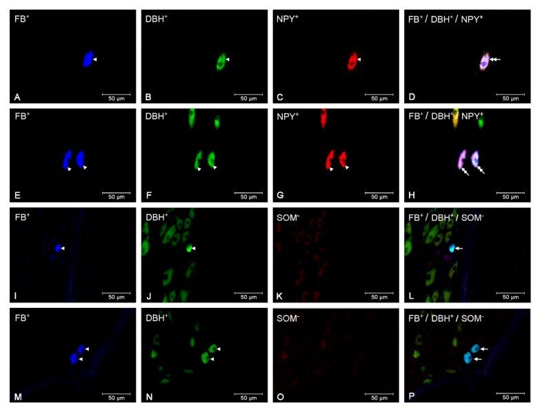
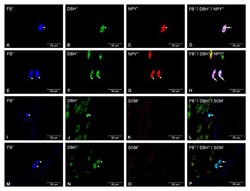
- Figure 3 Micrographs demonstrating the presence of DbetaH (B, F, J, N), NPY (C, G) and SOM (K, O) in the CaMG uterine perikarya of gilts from the CON ( A - D ), SAL ( I - L ) and E. coli ( E - H , M - P ) groups. The arrowhead indicates: Fast Blue (FB)-positive perikaryon, perikaryon presenting positive staining for DbetaH and NPY and perikaryon presenting positive staining for DbetaH. The double arrow indicates FB-positive uterine perikaryon expressing DbetaH and NPY. The arrow indicates FB-positive uterine perikaryon expressing DbetaH. The photographs (D, H, L, P) were made by digital superimposition of three color channels: FB-positive (blue), DbetaH-positive (green) and NPY- or SOM-positive (red). One large uterine perikaryon DbetaH and NPY immunoreactive is visible in the gilt of the CON group ( A - D ). In the CaMG of E. coli group, an elevation in the population of large perikarya containing these substances is observed ( E - H ). One small perikaryon expressing DbetaH, but not SOM, is present in the ganglion of the SAL group ( I - L ). In the E. coli group, two small perikarya expressing DbetaH, but not SOM, are observed in the CaMG ( M - P ).